4D D-Live
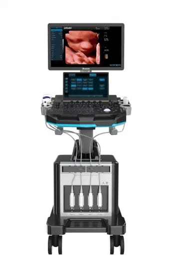
TROLLEY ULTRASONIC
DIAGNOSTIC APPARATUS
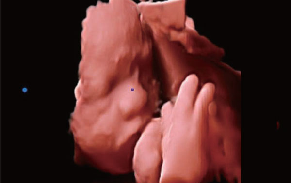
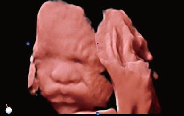
4D D-LIVE IMAGING
Beyond the limitations of traditional gray-scale ultrasound, the real-skin rendering is more intuitive, more three-dimensional, and more realistic than the four-dimensional color Doppler ultra- sound, and can record every move of the fetus into a video, and present it on the monitor for real-time viewing and sharing, creating the first “movie” for baby. 4D D-Live perfects well in detecting fetal abnormalities, the observation range is wider, and the picture is clearer.

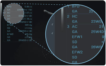
Professional OB & GYN
measurement packages
- Covering BPD, GS, EFW, EDD, GA, AFI, uterus, uterine appendage etc
- Obstetrics weight measurement formula: 13 kinds for option

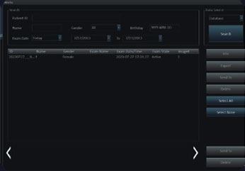
Smooth workflow, Easy management


TECHNOLOGY UPGRADE FOR CLEARER IMAGES
Digital beam enhancer
Analytical enhancement of the signal in the area by intelligent data to increase the clarity of the image
Multifold beam synthesis method
The clarity of the image is again enhanced by the overall technique of generating different multiple meta-beams in different forms, which are amplified by low noise and then output by the corresponding RF transceiver as bit baseband digits to cover a specific airspace to determine the target coordinates.
Excellent Imaging Processing Functions

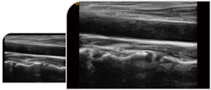
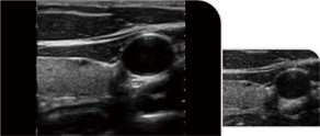
Trapezoid imaging is a kind of expanded imaging, which is transformed into a trapezoid based on the original rectangle, and the left and right sides are expanded to a certain extent, achieving a wider field of view the principle of ultrasound imaging is to scan the human body with ultrasonic sound beams, and obtain images of internal organs by receiving and processing the reflected signals.

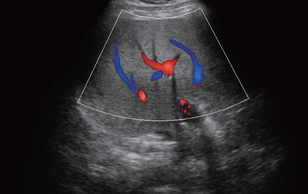
Directional Power Doppler employs a small sample volume with high resolution to produce images with two-color directional information and less ‘blooming’ of color for more realistic representation of defect size. It can make up for the lack Of power Doppler that can not display the direction of blood flow, and increase the direction information.

It improves image Clarity by improving tissue Contrast resolution, and spatial resolution, and eliminating near-field artifacts. It is mainly used for the diagnosis of cardiovascular and abdominal diseases. It plays an import ant role in evaluating the lesion area and boundary division of patients with imaging difficulties.
It can filter and extract the effective information of the whole frequency band and different depths, calculate the variation degree of the signal during the propagation process, perform targeted correction and matching, effectively suppress and filter the noise signal, and obtain high restoration imagings.
Ultrasound spatial composite imaging can improve contrast resolution, fine resolution and spatial resolution; enhance echo continuity at the tissue and lesion interface and reduce various artefacts (specular reflection, speckle, scatter, attenuation, poor contrast).

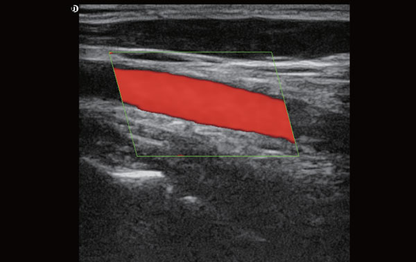
Triplex imaging is a combination of the Doppler, 2D image and spectral or pulse wave Doppler. The spectral Doppler helps to evaluate the velocity of blood flow. It is commonly used in arterial studies such as deep vein thrombosis scans to exclude a thrombus in a vein or in the carotid artery ultrasound scans.


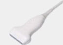
Showing Optional Probes
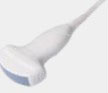
Abdomen, obstetrics, gynecology

Obstetrics, gynecology, urology

Fetal
Vascular, Musculoskeletal
Heart and chambers, cardiac function, pericardia, effusion

Baby organs

Prostate gland