
Exploring the vivid reality

Exclusive for Women’s Health
The P5 with 4D D-Live technology offers a com- plete solution that helps you with diagnostic confidence, efficiency and standardization in various diagnostic challenges in women’s health practice.

@ RealSkin reconstruction
Realskin rendering uses 4D ultrasound images and spatial dimensional parameters to obtain stereo-realistic 4D images that surpass most of the limitations of traditional grayscale ultrasound. Compared with 4D ultrasound, Realskin rendering is more complete in terms of the function of the detector, with a wider observation range and a clearer image.
@ Quick angle
Support 00, 900, 1800, 2700 rotation of 3D window
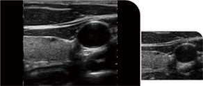
OB&GYN 4D D-Live Imaging
4D imaging, also known as real-time 3D imaging, provides an interactive means of viewing dynamic 3D imaging. 4D imaging examinations are performed with the volumetric probe fixed in one position and cannot be moved; the mechanical components inside the probe are able to perform stable continuous scanning of different positions by oscillation, resulting in a series of continuous and stable frame images. While D-live tech adopts a uinique and movable light source inside, providing impeccable views of baby’s organs.
Professional Measuring Packages
Professional obstetrics and gynecology measurement package:
Biparietal diameter, gestational sac, fetal weight, gestational period, gestational week, amniotic fluid index, uterus, uterine adnexa, etc.

Comprehensive other measurement package:
Specialised measurements available (such as distance, area, angle, time, slope, heart rate, velocity, acceleration, blood flow trajectory, blood flow spectral tracing, resistance index/beat index, etc.)

Smooth Workflow, Smart Interaction


Thin and Flexible Body

- 15″ full-view medical HD display
- 0-30 0 optimum angle adjustment
- All-in-one keyboard for easy operation


4 USB ports, 1 HDMI, 1 DP, 1 Audio, 2 RJ-45


Super Lightweight for Portablity
- Weight 9.0kg, standard with traveling case. Easy to Use in Every Possible Clinical Scenario:
outpatient services, emergency ambulance service, ICU, emergency room, point of care, etc. - 128GB SSD, Large storage for video images
- Built-in lithium battery, battery runtime > 2h
Exceptional Imaging Processing Functions

Trapezoidal Imaging
Trapezoid imaging is a kind of expanded imaging, which is transformed into a trapezoid based on the original rectangle, and the left and right sides are expanded to a certain extent, achieving a wider field of view. The principle of ultrasound imaging is to scan the human body with ultrasonic sound beams, and obtain images of internal organs by receiving and processing the reflected signals.
Spatial composite imaging
Ultrasound spatial composite imaging can improve contrast resolution, fine resolution and spatial resolution; enhance echo continuity at the tissue and lesion interface and reduce various artefacts (specular reflection, speckle, scatter, attenuation, poor contrast).

Tissue Harmonic Imaging (THI)
It improves image clarity by improving tissue contrast resolution, and spatial resolution, and eliminating near-field artifacts. It is mainly used for the diagnosis of cardiovascular and abdominal diseases. It plays an important role in evaluating the lesion area and boundary division of patients with imaging difficulties.
Micron Imaging
Micron imaging technology, real-time tracking of specific signals at the edges of different tissues, to achieve edge enhancement, and monitor each pixel at the same time; optimize the internal signal of the organization and perfectly integrate the edge info rmation and the internal pixel information of the organization to restore the real and delicate, excellent level contrast Two-dimensional image.

Doppler spectrum auto-envelope
Automatic and accurate envelope spectroscopy, automatic pressure, heart rate, resistance index and other haemodynamic indicators for accurate assessment.

Multiple Probes Optional
The self-developed probes with new array design technology and unique sub-array technology fully demonstrate the high resolution images from high density probes, perfectly presenting image details and increasing the accuracy of clinical diagnosis.
Abdomen, obstetrics, gynecology

Obstetrics, gynecology, urology

Fetal
Vascular, Musculoskeletal
Heart and chambers, cardiac function, pericardia, effusion

Baby organs

Prostate gland


