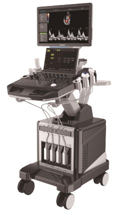
FULL APPLICATION COLOR
ULTRASOUNIC DIAGNOSTIC APPARATUS
Cardiology
DW-T5 Pro is a high-end color Doppler ultrasound that integrates multiple
technologies and performances excellently in cardiac ultrasound.
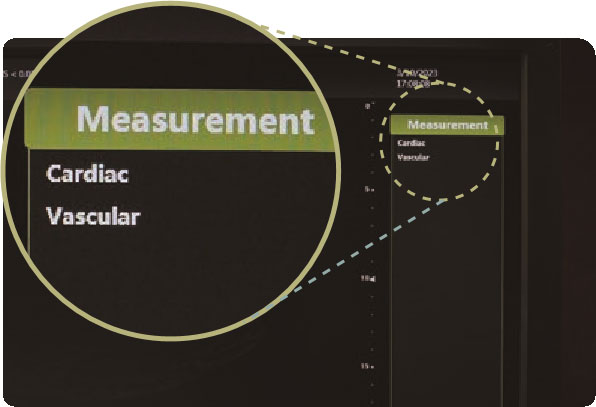
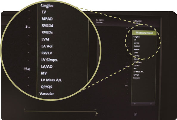
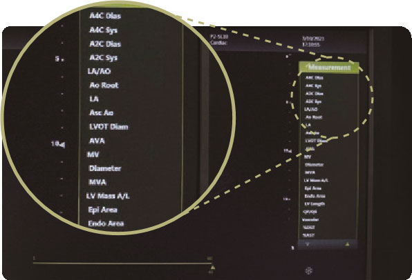
Specialist Measurement
Comprehensive Cardiac measurement package, covering all information of doctor’s diagnosis. Such as: aortopulmonary artery inner diameter, left ventricular myocardial performance index, etc.

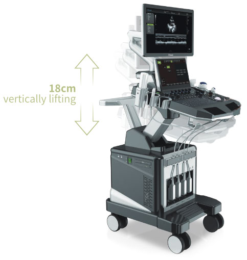
Unbelievable Design


Innovatice Platform

ST-U Technology Platform
- CPU+GPU dual-core processing architecture
- Clearer imaging, faster response
High-end Probes
- A/D conversion system+beam reconstruction technology
- Higher image resolution, more accurate clinical diagnosis
Meeting Your Needs
Real-time, manual, etc. optional modes

Optimizeing the angle of measurement automatically

Enhance biopsy needle visibility
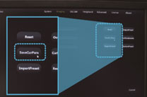
SSD 128GB+ HDD ITB:
Fast and stable startup

Ready to connect with PACS of hospitals
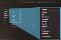
ZH/EN/VI/DE/FR/ES/RU/AR/PT/ID
Reducing duplication adjustment
Advanced Technology

FHI is a New harmonic imaging technology to reduce noise through the optimized filter, for automatically detecting the contour and getting a clearer image.
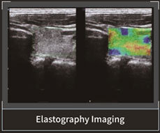
It uses the doppler principle to estimate tissue motion to obtain motion informa- tion and generate color-coded images of tissue motion speed.
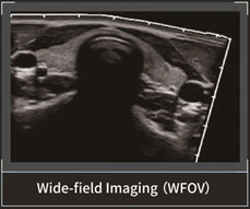
It is Omniview technology, which is not restricted by three orthogonal planes, the technology of artificially cutting volume data in any direction and angle through straight lines, curves, polylines, and trajec- tories of any plane or angle, so as to obtain any imaginary plane.

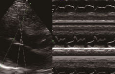
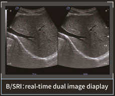
Speckle Reduction Imaging is a new algorithm that improves the image quality Of a-mode scanning by reducing reverber- ation artifacts. It helps heighten the visibil- ity of organs and lesions by suppressing noise.

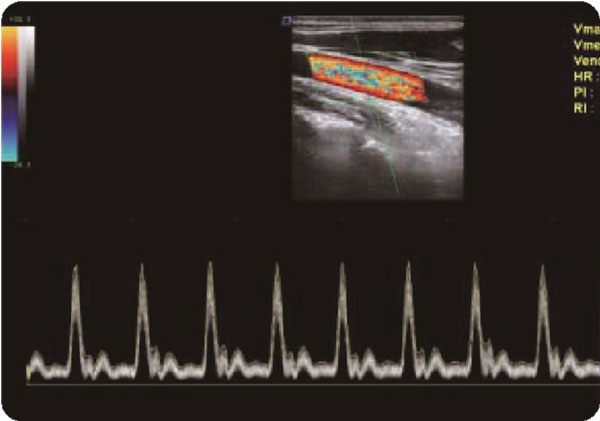
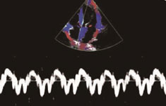
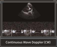
It is used for cardiac examination. Color blood flow uses speed and variance color maps to make colors superimposed on M mode images. Color blood flow covers a-mode images and M-mode timeline.
Technology It can enhance the signal of the tissue boundary through the deflection of the sound beam, reduces the phenomenon of side wall echo loss , and makes the bound- ary of the tissue clearer.


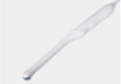
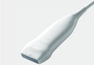
Showing Optional Probes
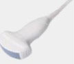
Abdomen, obstetrics, gynecology
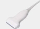
Vascular, Musculoskeletal
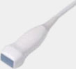
Heart and chambers, cardiac function, pericardia, effusion

Fetal
Obstetrics, gynecology, urology

Prostate gland

Baby organs
probe
Interventional ultrasound
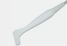
Interventional ultrasound
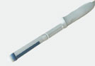
Andrology