
4D D-Live
TROLLEY COLOR ULTRASONIC
DIAGNOSTIC APPARATUS
General Imaging
Designed to help clinicians address the needs of diagnosis different body parts, DW-T5 Lite delivers crystal clear imaging capabilities.
Clinical Applications
- Abdomen
- Gynecology
- Obstetrics
- Pediatrics
- Small parts
- Vascular
- Cardiology
- Urology
- MSK
Professional Measuring Packages
DW-T5 Lite possess many speacialized measurement package of abdomen, obstetrics, urology, etc. for giving clinicians more scientific diagnosis assistent.


SIMPLIFING YOUR OPERATION


Accelerating Your Workflow

Advanced YH+ Operation Platform
- Matching high-speed 4G+128G Intel PC platform
- Improving data processing capabilities
- Getting Better man-machine interraction and inspection fluency

Sub-array Technology
- Fully displaying the high-resolution images Perfectly presenting image details
- Greatly increase the accuracy of clinical diagnosis
Meeting Your Needs

– Sharing Video and pictures on the PC side in real time via LAN

– Connecting with the PACS of hospital
– Quick Transmission

– Obtaining better images in a flash
– Saving operating time

– PSV, EDV, RI, PI, S/D, ACC, HR, etc
– Analyzing various data automatically

– Preseting the condition of the best images
– Reducing duplication process
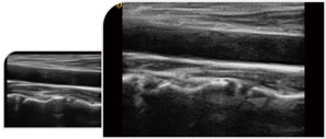
Giving You Better Imagings

Trapezoid imaging is a kind of expanded imaging, which is transformed into a trapezoid based on the original rectangle, and the left and right sides are expanded to a certain extent, achieving a wider field of view the principle of ultrasound imaging is to scan the human body with ultrasonic sound beams, and obtain images of internal organs by receiving and processing the reflected signals.

Directional Power Doppler employs a small sample volume with high resolution to produce images with two-color directional information and less ‘blooming’ of color for more realistic representation of defect size. It can make up for the lack Of power Doppler that can not display the direction of blood flow, and increase the direction information.

It improves image Clarity by improving tissue Contrast resolution, and spatial resolution, and eliminating near-field artifacts. It is mainly used for the diagnosis of cardiovascular and abdominal diseases. It plays an import ant role in evaluating the lesion area and boundary division of patients with imaging difficulties.

Micron imaging technology, real-time tracking of specific signals at the edges of different tissues. to achieve edge enhancement. and monitor each pixel at the same time; optimize the internal signal of the organization and perfectly integrate the edge information and the internal pixel information of the organization to restore the real and delicate, excellent level contrast Two-dimensional image.
It can filter and extract the effective information of the whole frequency band and different depths, calculate the variation degree of the signal during the propagation process, perform targeted correction and matching, effectively suppress and filter the noise signal, and obtain high restoration imagings.


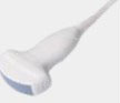
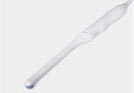
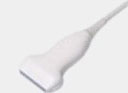
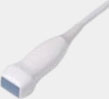
Showing Optional Probes
Abdomen, obstetrics, gynecology
Obstetrics, gynecology, urology

Fetal
Vascular, Musculoskeletal
Heart and chambers, cardiac function, pericardia, effusion

Baby organs

Prostate gland